Blood Vessels Labeled Brain / Blood Vessels Of The Brain Internet Stroke Center : Researchers have discovered how cells of the blood vessels sense the metabolic condition of the brain and alter vascular function in response.
Blood Vessels Labeled Brain / Blood Vessels Of The Brain Internet Stroke Center : Researchers have discovered how cells of the blood vessels sense the metabolic condition of the brain and alter vascular function in response.. Label the blood vessels of the male pelvis using the hints provided. Identify all of the blood vessels that are illustrated in the figure as you can while holding or otherwise examining whole brain specimens. Fill in the blanks with the appropriate words to describe blood flow from the heart. The central opening of a blood vessel, the lumen, is surrounded by a wall consisting of three layers: Posterior communicating a internal carotid а.
Blood supply to the brain is supplied by two main pairs of arteries, the internal carotid arteries and the vertebral arteries. Blood vessels in red in close communication with proliferating neuronal cells in the mouse cortex at embryonic day 10. Blood is supplied to the brain through 2 major pairs of arteries. If one of the major vessels becomes blocked, it is possible for blood flow to come across the circle of willis and prevent brain damage. Identify all of the blood vessels that are illustrated in the figure as you can while holding or otherwise examining whole brain specimens.
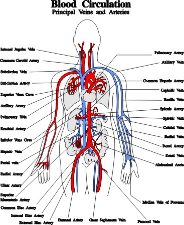
Blood Vessels Arteries Capillaries Veins Vena Cava Central Veins Lhsc from www.lhsc.on.ca Blood vessels are tubes that run through the transport system in which blood is transported. Blood vessel, a vessel in the human or animal body in which blood circulates. Supplies the anterior brain and the vertebral a. If there are blockages in blood vessels or if blood doesn't reach the brain for some reason, for how long can the brain maintain itself without oxygen? Veins are vessels that return blood to the heart. Traditionally, pais has been explained as being caused by a blood clot forming within the ageing placenta, entering the fetal circulation, embolising across the patent foramen ovale, travelling into the left ventricle, into the ascending aorta and then one of the main three branches of the thoracic aorta. Another whole article within the blood vessels and csf section is dedicated to the cavernous sinus. The central opening of a blood vessel, the lumen, is surrounded by a wall consisting of three layers:
Identify all of the blood vessels that are illustrated in the figure as you can while holding or otherwise examining whole brain specimens.
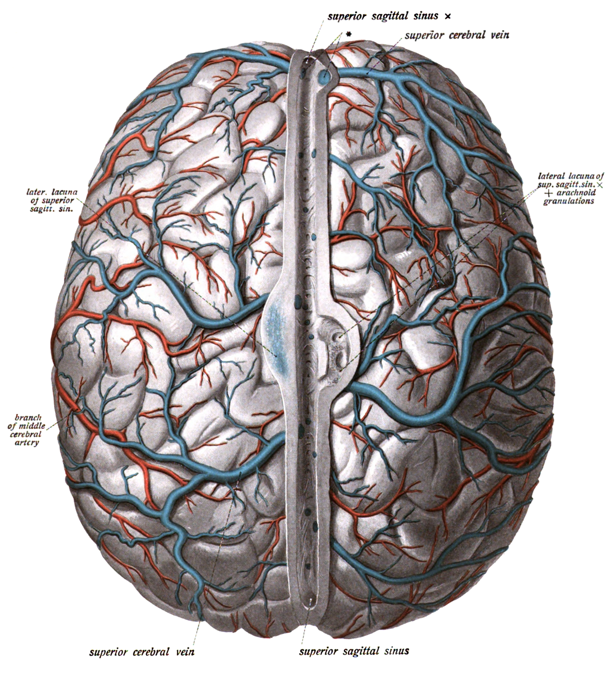
Comes off the subclavian a., ascends although the internal carotid a. The tunica intima is the inner layer facing the blood. The dense tight junctions between endothelial cells prevent paracellular transport through the. Supplies the anterior brain and the vertebral a. As well as providing new insights into the. What we see here are the blood vessels that extend along the inferior surface of the temporal and occipital lobe. Veins are vessels that return blood to the heart. The brain can autoregulate blood flow in order to ensure constant flow that is isolated from the arterial blood supply to the brain can be divided into the anterior and posterior circulation. Label the blood vessels in the inferior view of the brain using the hints provided. Only some of the vessels that exist in a real brain have been labeled. Fill in the blanks with the appropriate words to describe blood flow from the heart. These vessels transport blood cells, nutrients, and oxygen to the tissues of the body. The central opening of a blood vessel, the lumen, is surrounded by a wall consisting of three layers:
Only some of the vessels that exist in a real brain have been labeled. Cerebral arterial circle anterior communicating posterior cerebral a middle cerebral al reset zoom. They also take waste and carbon dioxide away from the tissues. The brain can autoregulate blood flow in order to ensure constant flow that is isolated from the arterial blood supply to the brain can be divided into the anterior and posterior circulation. Learn more about the anatomy and types of blood vessels and the diseases that affect them.
Superior Cerebral Veins Wikipedia from upload.wikimedia.org Blood is supplied to the brain through 2 major pairs of arteries. The brain can autoregulate blood flow in order to ensure constant flow that is isolated from the arterial blood supply to the brain can be divided into the anterior and posterior circulation. Label the blood vessels in the inferior view of the brain using the hints provided. Label the veins of the anterior forearm and hand. Blood supply to the brain is supplied by two main pairs of arteries, the internal carotid arteries and the vertebral arteries. Comes off the subclavian a., ascends although the internal carotid a. The central opening of a blood vessel, the lumen, is surrounded by a wall consisting of three layers: These vessels of the brain supply blood to the brain stem, the rear of the cerebrum and part of the cerebellum.
They do not have muscle layers and allow the exchange of substances between the blood and the a few structures (such as cartilage and the lens of the eye) do not contain blood vessels and are labeled avascular.
Function and homeostasis of the brain relies on communication between its complex network of cells. Identify all of the blood vessels that are illustrated in the figure as you can while holding or otherwise examining whole brain specimens. In the article on the ventricles within the cns, we will discuss their structure and. Only some of the vessels that exist in a real brain have been labeled. It is composed of an innermost layer of endothelium (simple squamous epithelium) surrounded by variable amounts of connective tissues. At the same time, blood vessel double labeled for brdu and reca were present in remote areas relative to the stroke lesion (figure 3h , arrows) and in the figure 4. Learn more about the anatomy and types of blood vessels and the diseases that affect them. If one of the major vessels becomes blocked, it is possible for blood flow to come across the circle of willis and prevent brain damage. The vessels that carry blood away from the heart are called arteries. These vessels of the brain supply blood to the brain stem, the rear of the cerebrum and part of the cerebellum. Blood is also supplied to the brain by the vertebral a. They do not have muscle layers and allow the exchange of substances between the blood and the a few structures (such as cartilage and the lens of the eye) do not contain blood vessels and are labeled avascular. The 500 ms patients, both adults and children, also underwent mri scans of the brain to measure iron deposits in surrounding areas of the brain.
The brain can autoregulate blood flow in order to ensure constant flow that is isolated from the arterial blood supply to the brain can be divided into the anterior and posterior circulation. Microscopically, it is formed by the endothelium of the blood vessel. The blood vessels (and nerves) enter the brain through holes in the skull called foramina. Blood is also supplied to the brain by the vertebral a. Blood supply to the brain is supplied by two main pairs of arteries, the internal carotid arteries and the vertebral arteries.
Circulation And The Central Nervous System Anatomy And Physiology I from s3-us-west-2.amazonaws.com Blood is also supplied to the brain by the vertebral a. Blood is supplied to the brain through 2 major pairs of arteries. They do not have muscle layers and allow the exchange of substances between the blood and the a few structures (such as cartilage and the lens of the eye) do not contain blood vessels and are labeled avascular. In the article on the ventricles within the cns, we will discuss their structure and. The vessels allow blood to be pumped at a high pressure to deliver nutrients and. Blood travels from the heart in arteries, which branch into smaller and smaller vessels, eventually becoming arterioles. Supplies the posterior brain, blood supply to the entire brain is ensured by anastomoses between the vessels. The blood vessels are the components of the circulatory system that transport blood throughout the human body.
Blood vessel, a vessel in the human or animal body in which blood circulates.
The vessels that carry blood away from the heart are called arteries. In the article on the ventricles within the cns, we will discuss their structure and. Blood travels from the heart in arteries, which branch into smaller and smaller vessels, eventually becoming arterioles. Label the veins of the anterior forearm and hand. Supplies the posterior brain, blood supply to the entire brain is ensured by anastomoses between the vessels. Blood vessels are tubes that run through the transport system in which blood is transported. Microscopically, it is formed by the endothelium of the blood vessel. The brain can autoregulate blood flow in order to ensure constant flow that is isolated from the arterial blood supply to the brain can be divided into the anterior and posterior circulation. Blood vessel endothelium is continuous with the inner tissue lining of organs such as the brain, lungs, skin, and heart. They also take waste and carbon dioxide away from the tissues. Blood vessels are intricate networks of hollow tubes that transport blood throughout the entire body so that it can deliver valuable nutrients to and remove waste from cells. Blood vessels are referred to collectively as the vascular system and, together with the heart, make up the circulatory system or cardiovascular system. Comes off the subclavian a., ascends although the internal carotid a.